
Angiography
Introduction
Angiography
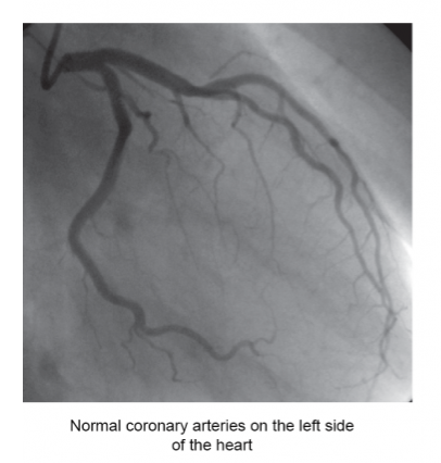
Angiography is an x-ray technique in which dye is injected into the chambers of your heart or the arteries that lead to your heart. Doctors can then measure the blood flow and blood pressure in the heart chambers and see if the coronary arteries are blocked. Doctors perform a cardiac catheterization procedure in which a long, thin tube (called a catheter) is put into an artery in the leg and threaded into the heart. Once the catheter is in place in the heart, a dye is injected through the catheter and into the heart. The dye helps doctors see how the heart chambers and the coronary arteries are working. The movement of the dye through your heart and coronary arteries is recorded as an angiogram and viewed on a television monitor.
Treatment
Risks associated with an Angiography:
- Small amount of radiation
- Extremely small chance you could develop cancer in the long term from the radiation
- If you have a known kidney disease
- Infection, bleeding or injury at the site of an injection
Why Angiograms are used?
Angiography is used to check the health of your blood vessels and how blood flows through them.
It can be used to help diagnose or investigate a number of problems affecting the blood vessels, including:
- atherosclerosis
- peripheral arterial disease (reduced blood supply to the leg muscles)
- a brain aneurysm (a bulge in a blood vessel in your brain)
- angina (chest pain that occurs when the blood supply to the heart muscle is restricted)
- blood clots or a pulmonary embolism
Benefits associated with an angiogram
- Used for diagnosis to show very detailed pictures of the arteries inside your brain, heart and kidneys
- Can be used to show blockages in your arteries